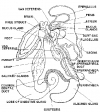
STAR FISH - GENERAL CHARACTERS
Monday, 05 June 2017 09:19Introduction
Star fish or Sea star is common echinoderm in the sea water. Its Zoological name is Asterias rubens. It belongs to,
- Phylum: Echinodermata
- Sub Phylum: Asterozoa
- Class: Stelleroidea
- Sub Class: Asteroidea
- Order: Forcipulata
The genus Asterias is represented by 100 species,
Eg: 1) A. rubens (North European sea coast), 2) A. vulgaris (North American sea coast)
- Star fish dwells on the bottom of the sea.
- It is a benthonic form.
- They are more common on hard rocky sea bottom, Star-fish is a carnivorous animal.
- It creeps slowly on the sea bottom.
- It can bend or twist in many ways.
- It has the power of autotomy (Self amputation).
External Features: The body of Asterias is star shaped and looks like a sea star.
Shape:
- The body, is compressed on oro-aboral axis.
- It shows a central disc. From this disc five arms will project.
- They are symmetrically arranged.
- The arms are board proximalhy and are free at the distal end.
- The arms occupy the radial axis.
Size:
- The smallest star fish is one cm in diameter, where as the largest one is 200 cm in diameter.
- The average size varies from 10 to 20 cm.
Colour:
Star fish show brilliant colours. Yellow - brown or orange colours are common.
Surface of the Body:
The body shows two surfaces.
- Oral and
- Aboral.
- The oral surface faces the bottom of the sea.
- The centre of the oral surface contains mouth.
- The aboral surface is directed upwards. It is slightly convex.
- The oral and aboral surfaces are not dorsal and ventral sides of the body, but right and left sides of a bilaterally symmetrical animal.
Oral surface or Actinal surface: It is the bwer surface of the animal. It is directed downwards. It is flat. It shows the following parts.
1. Mouth: It is round opening. It is present in the centre of the oral surface of the central disc. The mouth is also called Actinosome. It is surrounded by peristomial membrane, which is soft. Mouth is surrounded by five groups of oral spines.
2. Ambulacral grooves: From the comers of the mouth five ambulacral grooves will start and run along the middle of the arms.
3. Tube feet or Podia: Each ambulacral groove contains 4 rows of tube feet. They are soft and extensible tubular processes. Each tube feet end- in a sucker. The tube feet are useful for locomotion and food collection.
4. Ambulacral spines: Each ambulacral groove is guarded on each side by 2 or 3 rows of spines. These are movable. These spines are aggregated into five groups called Mouth papillae.
Three rows of stout immovable spines are present outside the ambulacral spines.
Another row of spines will present along the borders of the arms separating the oral from aboral surface.
5. Eye: The eye is small and bright red spot. It is present at the end of each arm. It is light sensitive.
6. Tentacle: At the end of each arm a small non-retractile tentacle is present. It is olfactory in function.
Aboral surface or Abactinal surface: The surface of the star firsh which is facing upwards is called aboral surface. It is convex. It has the following structures.
1. Anus: It is a small opening. It is nearly in the middle of aboral surface. It is slightly displaced towards the interradius.
2. Madreporite: It is flat, almost circular plate. It is sieve plate like structure. It leads into a stone - canal of water vascular system. Madreporite is placed in an inter-radius of the central disc. It converts the radial symmetry of the animal into bilateral symmetry.
3. Spines: The spines of aboral surface are white in colour. They are arranged in irregular radial rows. The spines are short, and stout. They are developed from the calcareous plates called ossicles. The ossicles are burried in the body wall and covered by epidermis.
4. Dermal branchiae or Papulae: These are small finger like processes. They come but through dermal pores. Dermal pores are present in between the ossicles. These papulae are also called gills. They are respiratory in function. They can also perform excretory function. These papulae can extend or completely retracted into the body.
Pedicellariae: Pedicellariae are scattered all over the body. These are present in between the spines of the aboral surface. On the oral surface they are present attached to the bases of the spines.
Structure: The pedicellariae are stalked. They are called pedunculate type. Stalk is covered by epidermis which has sensory and gland cells.
It shows three calcareous ossicles.
1. Basilar piece near stalk.
Two jaws, the jaws are articulate with the basilar piece. They can be opened or cbsed like the jaws of a bird by muscles such pedicellariae, are called forclpulate.
Kinds: Two types of forcipulate pedunculate pedicellariae are seen in Asterias. In the forceps or straight type, two basal ends of the two jaws are arranged like Forceps. In crossed or scissors type the two basal ends of the two jaws are crossed and look like scissors. The jaws are also operated by three pairs of muscles.
Functions:
- They protect the delicate gills or papulae.
- They remove the debris form body.
- They are helpful in the capture of small prey.

INTRODUCTION TO PHYLUM ECHINODERMATA
Monday, 05 June 2017 09:03
EXCRETORY SYSTEM IN UNIO
Monday, 05 June 2017 08:52
CHARACTERS AND CLASSIFICATION OF PHYLUM MOLLUSCA
Sunday, 04 June 2017 20:51
REPRODUCTIVE SYSTEM IN PILA (SNAIL)
Sunday, 04 June 2017 18:59
EXCRETORY SYSTEM IN PILA (SNAIL)
Sunday, 04 June 2017 18:22
BLOOD VASCULAR SYSTEM IN PILA (SNAIL)
Sunday, 04 June 2017 17:06
SENSE ORGANS IN PILA (SNAIL)
Sunday, 04 June 2017 08:43
NERVOUS SYSTEM OF PILA (SNAIL)
Sunday, 04 June 2017 08:10
RESPIRATORY SYSTEM OF PILA (SNAIL)
Sunday, 04 June 2017 07:31
DIGESTIVE SYSTEM AND PROCESS OF DIGESTION IN PILA GLOBOSA (APPLE SNAIL)
Saturday, 03 June 2017 13:47
STRUCTURE, PALLIAL COMPLEX AND INTERNAL SOFT PARTS OF PILA GLOBOSA (APPLE SNAIL)
Saturday, 03 June 2017 12:57
PHYLUM MOLLUSCA
Friday, 02 June 2017 22:05


