Lichtman's Atlas of Hematology
- ISBN-10: ###
- ISBN-13: ###

Synopsis
Preface: One attraction of the clinical discipline of hematology has been the ability to diagnose blood cell disorders quickly and relatively simply using tests that can be performed…
0MB
Size
3402
Views
Preface: One attraction of the clinical discipline of hematology has been the ability to diagnose blood cell disorders quickly and relatively simply using tests that can be performed in a hematologist's office: notably, blood cell counts, blood films, the reticulocyte count, and film of the marrow aspirate. Now these procedures are often performed in clinical laboratories, most with automated techniques. Nevertheless a physician, experienced and competent in the interpretation of blood counts and blood film, can determine the diagnosis or can reach a very limited differential diagnosis, quickly, in many cases.
Findings on marrow film were the centerpiece for understanding the stages of precursor development of erythroid, myeloid, and megakaryocytic lineages and were essential elements in the determination of the cytokinetics of the blood precursor compartments. Findings on blood film have provided clues to the pathogenesis of disease, such as processes leading to hemolysis, including inherited and acquired membrane or hemoglobin abnormalities, and mechanical disruption of red cell integrity in the circulation or the microvasculature. Landmark work making the marrow and blood film useful was contributed by Paul Ehrlich, chemist, immunologist, and Nobel prize laureate, based on studies of aniline dyes, described in his publication, Farbenanolytische Untersuchungen zur Histologie und Klinik des Blutes, Berlin, 1891. This work provided the basis for the polychrome stains used to distinguish blood and marrow cells.
Today, blood cell counts and blood films are supplemented by immunophenotyping, cytochemistry, and immunostaining of blood, marrow, or lymph node cells, computerized performance of cytogenetic analysis, and gene expression profiling. Thus, we have included in this Atlas prototypical flow cytometric findings, cytochemical and immunostain reactions, and cytogenetic findings useful in the diagnosis of hematological diseases. A display of important external and cutaneous lesions is also included, the latter often the result of coagulant and anticoagulant protein abnormalities.
Despite the increased specificity of hematological diagnosis provided by important ancillary analytical techniques, in many cases the blood counts and the blood and marrow films still permit the physician, expert in hematology, to deduce much of the information required to reach a diagnosis and to plan therapy. This initial diagnostic consideration can be supplemented by the important, indeed sometimes vital, additional testing available. Nevertheless, the presence of polychromatophilic macrocytes (reticulocytes) and characteristic red cell shape changes on the blood film may point to the specific type of hemolytic anemia, the white cell count and the differential count may be diagnostic of one of several types of leukemia, the platelet prevalence and morphology may be indicative of any of several thrombocytic disorders, and innumerable other diseases may be detected with the blood or marrow film, e.g., myeloma, lymphoma, Gaucher disease, Chediak-Higashi disease, and many others. A number of infectious diseases may be evident by the organisms parasitizing red or white cells. Numerous such examples are represented in this Atlas.
The intent of this work is to provide on-line access to images that are characteristic of a wide array of hematological disorders and other disorders that affect the blood, enhancing written descriptions found in Williams Hematology and other relevant medical books published on-line by McGraw-Hill. This Atlas permits an extensive presentation of blood, marrow, lymph node, spleen, and other organ pathology that would be too costly to be included in the print version of the text.
Experience is required to use blood cell morphology accurately and wisely. Even the expert morphologist may have difficulty being certain about mean red cell size or hemoglobin content, given the requirement to estimate cell volume from the area of red cells on a dried film or the sometimes irregular distribution of hemoglobin in cells on a blood film. Moderate or marked changes are usually discernible but early, slight, or mild changes may be undetectable by the observer. Some macrocytes may be so-called “thin macrocytes” as in thalassemia, and have a low red cell volume, whereas oval macrocytes, characteristic of megaloblastic anemia are “thick macrocytes” and have a marked increase in cell volume, despite the area of each type of macrocyte on the blood film being similar. Nevertheless, the blood and marrow films remain an invaluable source of information. Indeed, in experienced hands, in many cases, much of what may be found in the marrow can be deduced on a careful analysis of the blood.
For the clinical microscopist, examination of blood and marrow stained films is an adventure in the search for a patient's diagnosis. The stained film can instill an appreciation for the remarkable effects pathological processes can produce on blood cells. The deviations in red cell morphology, viewed by scanning electron microscopy, have also led to artistic expression as displayed in Corpuscles: an atlas of blood cell shapes by Marcel Bessis published in 1974 by Springer-Verlag. Thus, the anticipated technical advancements allowing information from a blood or marrow film to be harvested by electronic machines or replaced by gene expression or protein expression patterns may increase the reproducibility and, perhaps, the accuracy of diagnosis but will result in the loss of an important and fascinating medical endeavor, cell microscopy. Cell microscopy has been vital to the knowledge gained by cell biologists over the last four centuries ever since the cloth merchant, Antony van Leeuwenhoek of Delft, as a result of his avocation, lens-making, first described blood corpuscles, previously thought by Marcello Malphigi to represent fat globules. The important relationship of blood, marrow, and lymph node cells to their environment (stroma) cannot be captured in an atlas of blood cells. In the case of cell biology, however, one must always consider the following admonition:
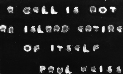
This quotation, from the noted biologist Paul Alfred Weiss, was formed by scanning electron micrographic images of misshapen red cells from a patient with thalassemia minor and a patient with sickle cell anemia.
Marshall A. Lichtman, M.D.
Rochester, NY, USA
2007
Lichtman's Atlas of Hematology
Download locations
0MB file size
All files are original, bioscience.com.pk does not repack or modify downloads in any way.
Trouble downloading? Try Internet Download Manager (IDM)
- Posted by Chetan Dg.
End of the synopsis