Displaying items by tag: Books
Clinical Parasitology: A Practical Approach
Description: Now in full color, the second edition of Clinical Parasitology provides you with all of the information needed to perform, read, and interpret parasitology tests in a clear and understandable way. The user-friendly design, extensive illustrations, pedagogical features and clear descriptions of look-a-like parasites will help you better hone your skills and confidently perform clinical procedures.
Obstetrics & Gynecology PreTest Self-Assessment & Review 12th Edition (PreTest Clinical Medicine)
Description: The student tested-and-reviewed way to prep for the Obstetrics and Gynecology shelf exam and the USMLE Step 2 CK Obstetrics & Gynecology: PreTest™ Self-Assessment & Review is the perfect way to assess your knowledge of obstetrics and gynecology for the USMLE Step 2 CK and shelf exams. You'll find 500 USMLE-style questions and answers that address the clerkship's core competencies along with detailed explanations of both correct and incorrect answers. All questions have been reviewed by students who recently passed the boards and completed their clerkship to ensure they match the style and difficulty level of the exam. 500 USMLE-style questions and answers Detailed explanations for right and wrong answers Targets what you really need to know for exam success Student tested and reviewed Obstetrics & Gynecology: PreTest™ Self-Assessment & Review is the closest you can get to seeing the test before you take it. Great for clerkship and the USMLE Step 2 CK! Obstetrics & Gynecology: PreTest asks the right questions so you'll know the right answers. Open it and start learning what's on the test.
Nephrology and Clinical Chemistry: The Essential Link
Description: Description: Clinical chemistry is a science that requires specific knowledge and teaching. The e-book covers several topics in clinical nephrology. Patients suffering from severe chronic kidney disease may be quasi asymptomatic. This lack of overt symptomatology suggests that clinical laboratory tests are of the highest importance. Clinicians treating patients and clinical biologists operating in laboratories are often far apart from each other. They have a lot of things to share and to learn from each other. This e-book bridges the gap between these two groups of health professionals.
Practical Management of Chronic Viral Hepatitis
Description: Continuous acquisition of new knowledge in Medicine is essential to ensure progression in diagnostics and therapeutics. In the last decade the discipline of Hepatology has achieved critical progress in the treatment of viral hepatitis. The present book has been realized by a team of experts daily facing clinical problems in the prevention and management of liver diseases and has been designed for a global readership to offer some practical tips to physicians who want update their level of practice in the field. Its a practical volume for daily reference but also an instrument for improving expertise in viral hepatology and discovering the unresolved issues. Management of HBV and HCV hepatitis in young and elderly, HEV hepatitis, evaluation of liver fibrosis, hepatocellular carcinoma, vaccine and prevention and patient education are some of the most important topics covered by the authors. In addition, an outstanding chapter on the skin involvement during viral hepatitis and the tools to manage them during triple therapy is included in the book.
Pathology Mini Tutorials University Of Nottingham
Description: Short tutorials illustrated with gross and microscopic photographs of pathological conditions. These video podcasts are designed to supplement pathology tutorials for Nottingham medical students.
Diagnostic Pathology and Molecular Genetics of the Thyroid
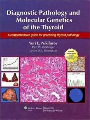
Description: The first comprehensive surgical pathology textbook and reference on the thyroid in over fifteen years, this book presents the most advanced concepts on the diagnostic surgical pathology, cytopathology, immunohistochemistry, and molecular genetics of neoplastic and non-neoplastic thyroid diseases. The authors provide a detailed description of the surgical pathology of thyroid diseases side-by-side with major advances in immunohistochemistry and molecular genetics that can be used in evaluating thyroid tumors and non-neoplastic diseases. By combining diagnostic surgical pathology, cytopathology, immunohistochemistry, and molecular genetics, the book effectively mimics the practice of contemporary surgical pathologists. All major chapters have a uniform style of description and include a separate section on molecular genetics.
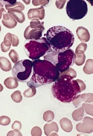
Lichtman's Atlas of Hematology
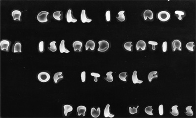
This quotation, from the noted biologist Paul Alfred Weiss, was formed by scanning electron micrographic images of misshapen red cells from a patient with thalassemia minor and a patient with sickle cell anemia.
Rare Hematological Malignancies
Description: This hugely practical work will be a bible in the pocket of hematologists and other practitioners everywhere, covering as it does malignant hematologic diseases that physicians will only occasionally see. It provides accurate, up-to-date information on the disease biology as well as practical recommendations concerning disease management. Information concerning these diseases, and particularly regarding their management, can be extremely difficult to find. Not any more.
Diagnostic Cytopathology by Gray
Description: * 2011 BMA Book Awards - Highly Commended in Pathology * The new edition of Diagnostic Cytopathology provides the practicing and trainee cytopathologist with a comprehensive guide to the diagnostic applications of exfoliative and aspiration cytology. The book covers normal and abnormal cytological findings encountered in all body sites where cytological applications are used. Appropriate histopathological, immunohistochemical and molecular correlations, together with a consideration of the possible differential diagnoses, accompany the cytological findings. The reader can see a full range of normal and abnormal findings with almost 2,000 full-colour images. The book is heavily referenced to ensure that it will serve as a practical resource for daily reference in the laboratory. A uniformity of basic chapter structure will help readers to quickly find the diagnostic answers they seek. A practical in-depth bench book that covers everyday diagnostic work in the laboratory. It provides an accessible guide to cytological diagnostic investigation and screening. Each chapter provides a summary of major diagnostic criteria in order to quickly direct the user to the most relevant material. Cytological findings closely related to histopathological, immunohistochemical and molecular appearance whenever appropriate to assist in the interpretation and recognition of tissue samples. Almost 2,000 colour illustrations incorporated throughout to provide a comprehensive visual guide. Stay on top of the latest concepts and developments in molecular markers. Access full text online and download images via Expert Consult. This edition stresses not just the diagnostic cytological features of the various conditions encountered, but also the diagnostic pitfalls and the grey areas between so as to enable the reader to give more evidence-based reports. In recognition of their rapid expansion, there are new chapters on recent technological developments and on the cytodiagnosis of childhood tumours. A special section on the importance of multidisciplinary team meetings that include the cytopathologist as a core member of the team has also been included at the end of each chapter. As active members of this team, cytopathologists can define their role in the management pathway and thus bring the patient and the microscope together as never before. The full text can be accessed online and images downloaded via Expert Consult.
Papillomavirus Infections in Human Pathology
Description: This is the most up-to-date and comprehensive reference source on papillomavirus-associated human pathology, which examines the molecular biology of the infections and discusses diagnosis and treatment methods. Abundantly illustrated, it covers all aspects of human papillomaviruses from molecular biology and pathogenesis of HPV-associated diseases, as well as natural history and epidemiology, to a detailed study of the role of these viruses as potential etiological agents of a wide spectrum of both benign and malignant human tumours. While the main focus is on HPV infections of the genital tract, lesions of the skin, respiratory tract, the digestive system, the urinary tract and the eye are also covered in detail. Written by two authors who are internationally renowned for their work, it will be invaluable for both clinicians and researchers working in the fields of gynecology, pathology, sexually transmitted diseases, genito-urinary medicine, oncology, dermatology, otorhinolaryngology, and cancer epidemiology.
English for Science by Fran Zimmerman
Description: A text-workbook for use in secondary school and higher levels preparing ESL students for basic science courses taught in English.
Difficult Diagnoses in Breast Pathology
Description: Description: Breast cancer is the second leading cause of cancer death in women in the United States. For the pathologist, almost any breast lesion may produce diagnostic difficulty, especially due to frequently small samples (core biopsy specimens) and a variety of mimics and variants seen in specific types of lesions. Additionally, the difficulty of breast lesion diagnosis has risen dramatically in recent years due to the increased emphasis on stratifying patients for appropriate therapy on an individual basis; the wider range of both local and systemic therapeutic options, and the potential for earlier diagnosis through increased mammographic breast screening leading to a higher likelihood of a favorable outcome. Difficult Diagnoses in Breast Pathology provides a highly visual presentation of the major problems and questions that a pathologist is likely to encounter in the evaluation of common and uncommon breast diseases. Coverage includes needle core biopsy interpretation, diagnosis of precursor lesions, early stage disease, and recognition of neoplastic mimics and other misleading variants. In addition, this book emphasizes particularly difficult areas including the use of newer immunohistochemical markers. Throughout, the emphasis is on an easily accessible presentation with tables and lists of key points summarizing major findings and numerous high-quality images supporting the text. Difficult Diagnoses in Breast Pathology will be a valuable reference for every pathologist who deals with the diagnosis of breast diseases.
Atlas of Hematopathology: Morphology, Immunophenotype, Cytogenetics, and Molecular Approaches
Description: As the definitive diagnostic atlas of the diseases of the hematopoietic system, the Atlas of Hematopathology appeals to a wide range of people who are being trained in a variety of medical fields or practicing as non-hematopathologists, and therefore, are looking for a book which can provide information in a clear, focused format, with no excessive text or details. The atlas offers effective guidance in evaluating specimens from the lymph nodes, bone marrow, spleen, and peripheral blood, enabling clinicians to deliver more accurate and actionable pathology reports. Practicing physicians and those in pathology and hematology training also gain a better understanding of the nature of hematologic disorders and improve their diagnostic skills along the way. Taking a unique multi-disciplinary approach, the book covers conventional histopathology and cytopathology, as well as all important complementary diagnostic tests, such as immunophenotyping (immunohistochemical stains and flow cytometry), karyotyping, FISH and DNA/molecular studies. It offers concise textual and extensive visual coverage of both neoplastic and non-neoplastic hematology disorders, with the neoplastic hematology sections presented according to the most recent WHO classifications. There is also an introduction to the normal structures of hematopoietic tissues and the various multidisciplinary techniques. The atlas contains more than 900 high-quality color images that mirror the findings that fellows and clinicians encounter in practice. It provides information in a quick, simple and user-friendly manner, attracting those who are in training or are not considered experts in the field. Residents, fellows, practicing clinicians, and researchers in pathology, hematology, hematology/oncology, as well as graduate students in pathology and other clinicians workings in clinical hematology laboratories will all find it useful. Saves clinicians and researchers time in quickly accessing the very latest details on the diverse clinical and scientific aspects of hematopathology, as opposed to searching through thousands of journal articles For clinicians, fellows, and residents, correct diagnosis (and therefore correct treatment) of diseases depends on a strong understanding of the molecular basis for the disease - hematologists, pathologists, oncologists, and other clinicians will benefit from this clear, focused, annotated format Companion web site features over 900 images from the book!
Diagnostic Histopathology of Tumors by Fletcher
Description: Diagnose tumors with confidence with Diagnostic Histopathology of Tumors, 4th Edition. Dr. Christopher Fletcher's renowned reference provides the advanced, expert guidance you need to evaluate and interpret even the most challenging histopathology specimens more quickly and accurately. Diagnose efficiently and effectively using diagnostic flow charts, correlations of gross appearances to microscopic findings, and differential diagnosis tables for better recognition and evaluation of similar-looking entities. Employ immunohistochemistry, molecular and genetic diagnostic tests, and other modern techniques as well as the best morphologic diagnostic methods to effectively identify each tumor or tumor-like entity. Utilize new, clinically important molecular genetic data and updated classification schemes to help guide treatment and targeted therapy. Apply the latest techniques and diagnostic criteria with completely rewritten chapters on Small and Large Intestines, Heart, Larynx and Trachea, Ear, and Peritoneum. Find critical information quickly thanks to more tables and bulleted lists throughout. Access the entire text and illustrations online, fully searchable, at www.expertconsult.com.
Underwood's Pathology: A Clinical Approach
Description: Underwood's Pathology (formerly General and Systematic Pathology) is an internationally popular and highly acclaimed textbook, written and designed principally for students of medicine and the related health sciences. Pathology is presented in the context of modern cellular and molecular biology and contemporary clinical practice. After a clear introduction to basic principles, it provides comprehensive coverage of disease mechanisms and the pathology of specific disorders ordered by body system. An unrivalled collection of clinical photographs, histopathology images and graphics complement the clear, concise text. For this sixth edition, the entire book has been revised and updated. Well liked features to assist problem-based learning - including body diagrams annotated with signs, symptoms and diseases and a separate index of common clinical problems - have been retained and refereshed. Additional value is provided by the complementary online version - hosted on studentconsult.com - which includes the complete, fully searchable text, downloadable images, clinical case studies and a revised, interactive self-assessment section to check your understanding and aid exam preparation. This all combines to make Underwood's an unsurpassed learning package in this fascinating and most central medical specialty.
Atlas of Acoustic Neurinoma Microsurgery
Description: This colour atlas provides information on all major acoustic neurinoma procedures, and is designed to improve physicians’ skills in this specialty. With more than 50,000 new cases per year worldwide, acoustic neurinomas are among the most commonly observed skull-base tumours. They are also among the most demanding for surgeons, since great precision is needed to safeguard the auditory nerve, the facial nerve and other critical structures. The book begins with sections on the radiographic assessment of tumours, the selection of surgical approach, and pre-operative preparations. It then turns to the main surgical approaches, using hundreds of photographs-taken during actual surgery-to lead readers through each procedure in step-by-step detail. There is a special section on the use of endoscopy in acoustic neurinoma surgery, and an analysis of 50 MR scans. This practical volume should be of interest to otolaryngologists, neurosurgeons and other physicians involved in acoustic neurinoma surgery.